Pages Menu

Posted on Jun 3, 2014 in Conference | 0 comments

Socially Optimized Learning in Virtual Environments (SOLVE): Developing, Evaluating, and Disseminating A Game HIV Prevention Intervention Nationally Over the Web


Lynn C. Miller, PhD1, John L. Christensen2, Paul Robert Appleby3, Stephen John Read3, Stacy Marsella3, Charisse Corsbie-Massay4, Carlos Godoy3, Mei Si5, Janeane Anderson3, David Jeong3, Mina Park3

1Professor, USC Annenberg School for Communication and Journalism & Department of Psychology; 2University of Connecticut; 3University of Southern California; 4Syracuse University; 5Rensselaer Polytechnic Institute

Journal MTM 3:1S:4, 2014

DOI: 10.7309/jmtm.3.1S.2

Abstract


Young men (18–24) who have sex with men (YMSM) are at high risk for contracting HIV. Most existing HIV prevention interventions focus on changing intervening cognitive and deliberative processes or outcomes (e.g., beliefs, norms, self-efficacy, intentions) to change behavior. Many MSM, however, guided by contextual cues in emotionally arousing scenarios, make more automatic risky decisions they later regret. One emotion in a sexual narrative that might precipitate more automatic risky choices for young MSM may be shame (e.g., in one’s sexual desires). But, HIV prevention interventions are not designed to reduce MSM’s shame. SOLVE (Socially Optimized Learning in Virtual Environments), as demonstrated by an NIAID-funded randomized control trial, used a sex-positive game to reduce MSM’s shame, increase traditional immediate cognitive outcomes, and reduce unprotected anal intercourse for young Black, Latino, and White MSM (18–24) over 3 months. Could interactive interventions be delivered more broadly over the web? In prior CHRP funded work, a SOLVE interactive video (IAV) intervention was streamed over the web throughout California. However, an IAV approach limits the amount of user interaction, risk challenges users receive, and intervention tailoring to MSM’s decisions. This is addressed using a nationally deliverable 3D animated intelligent agents/interactive digital storytelling game in UNITY. MSM design their own characters, make choices for them on dates and sexual interactions, and are scaffolded by the user character’s virtual future self (participant’s older chosen self-character) to enhance self-regulation when risky. The NIMH-funded SOLVE-IT game development process for young MSM is discussed. Preliminary results from a 6-month randomized controlled trial conducted nationally, over the web, are promising.


*A similar abstract was published in a previous version of your journal.

Read More

Posted on Jun 3, 2014 in Conference | 0 comments

Innovative Use of Technology for HIV Prevention and Care: Evidence, Challenges and the Way Forward


Sean Young, PhD, MS1, Jason Chiu, MS1

1Department of Family Medicine, University of California, Los Angeles

Journal MTM 3:1S:1–3, 2014

DOI: 10.7309/jmtm.3.1S.1

Abstract


Use of mobile technologies for HIV prevention and care is no longer a promise, but a reality. From 2006 to 2009, a simple search of technology-related HIV studies yielded 1,147 results on PubMed and 274 results on PsycINFO. The number of technology-related HIV studies grew substantially in the next four years, as from 2010 to 2013 there were 1629 on PubMed and 407 studies on PsycINFO. These findings suggest a surge of interest in the field of technology use in HIV prevention and care research.

In response to this growth, the Center for HIV Identification, Prevention, and Treatment Services (CHIPTS) at the University of California, Los Angeles (UCLA) hosted the “Innovative Use of Technology for HIV Prevention and Care: Evidence, Challenges and the Way Forward” conference on January 23rd, 2014. Bringing together interdisciplinary researchers, community practitioners, clinicians, policy makers, technologists, and representatives from governmental and funding agencies, the conference sought to establish a collaborative framework for individuals to share their knowledge and experience in technology-based HIV prevention and care with each other.

Although the conference included information on different types of technologies, there was a particular emphasis on the role of mobile technology in HIV prevention and care. Popular mobile technologies are typically described as belonging to one or more the following categories: 1) cell phones, 2) native and cloud-based applications, 3) social media, and 4) mobile websites. Individuals differ in the way they use mobile technologies to communicate with each other, making it important to study the various ways in which mobile technologies can be used for HIV prevention and care. For example, cell phones facilitate multiple forms of communication, such as phone calls, short message services (SMS)/texts, and multimedia messaging services (e.g. video and picture texts)1. In the U.S., Youth (ages 18 to 29) communicate with each other predominately through SMS2, and recent HIV interventions targeting youth have utilized SMS to reduce substance use and increase HIV testing3,4.

The recent explosion in the use of social media can have a tremendous impact on HIV research, because these technologies can serve as platforms not only to reach a large number of at risk individuals but also to gather data on the behaviors of these individuals. Social media is defined as technologies, platforms, and services that enable individuals to engage in communication from one-to-one, one-to-many, and many-to-many5. In 2013, 73% of adults and 80% of teens used some form of social media6,7. There are many different types of social media, including 1) social networking sites (e.g. Facebook and MySpace), 2) blogs (e.g. Word Press or Tumblr), 3) microblogs of real-time communication (e.g. Twitter), 4) forum/bulletin boards (e.g. WebMD and Yahoo Answers), 5) social games (e.g. FarmVille and Mafia Wars), 6) media sharing (e.g. YouTube and Instagram), and 7) geosocial networking (e.g. Grindr and Jack’d). There exist different patterns of social media use based on socioeconomic, regional, and language factors, and before incorporating these technologies, researchers should understand these trends and how they impact HIV risk. For example, in the U.S., racial/ethnic (African Americans and Latinos) and sexual (e.g. gay and bisexual) minority individuals were found to be the most avid social media users8,9. Moreover, many African American and Latino men who have sex with men (MSM) have also used online social networks/geosocial networking apps to meet sexual partners to avoid potential stigma10,11. Taking into account information on patterns of social media use is important in learning how to craft HIV interventions and studies using social media among at-risk groups.

This conference served as an opportunity to introduce individuals to the latest mobile and social media technologies, and demonstrate research on how technologies can be used to study and address HIV among at-risk communities, along with a focus on the ethical and business-related issues associated with these approaches. The objectives of the conference were: 1) to present the most recent research findings and trends in HIV/AIDS and technology, 2) to increase the capacity of participants to develop, to implement, and to evaluate effective and evidence-based HIV/AIDS interventions with technology in clinical, research and community settings, and 3) to bring together researchers, clinicians, and technologists and to foster interdisciplinary collaborations on innovative way to improve HIV/AIDS prevention among at-risk communities.

The following topics emerged from the conference that can be used as a reference for the current state of the field, including research, community-based practice, and ethical considerations: 1) Social media and mobile technologies are increasingly being used by HIV researchers: A number of presentations focused on the use of mobile technologies, including using them as methods for recruitment, interventions, and data collection. Because of the increasing use of social media among populations at risk for HIV and highly-affected by HIV (e.g., minority MSM), researchers should continue exploring innovative ways to make use of these technologies for HIV prevention and care. 2) Community-based organizations and clinics are receptive and interested in applying social media and mobile technologies to HIV prevention and care: We heard from clinic senior management who described their interest and enthusiasm in using mobile technologies to address disparities in HIV prevention and care. Local organizations are interested and willing to partner with academic researchers on this work. 3) The ethical considerations around using these technologies for HIV prevention and care are only beginning to be explored: Our ethics panel conversation was focused primarily on a mobile application related to individuals affected by HIV and the ethical concerns of using mobile applications for HIV prevention and care. It was an initial step in providing a conversation on this needed topic and highlighted the interest and importance in establishing guidelines12 for safety and confidentiality in technologies that might increase HIV risk, as well as those that are used for HIV prevention and care. This special issue of the journal is dedicated to providing the readers with the abstracts from the presenters of the conference.

References

1. Maximizing Mobile. Washington D.C.: The World Bank, 2012.

2. Duggan M. Cell Phone Activities 2013. PEW Research Center, 2013.

3. Reback C, Grant D, Fletcher J, Branson C, Shoptaw S, Bowers J, et al. Text Messaging Reduces HIV Risk Behaviors Among Methamphetamine-Using Men Who Have Sex with Men. AIDS and behavior. 2012 2012/10/01;16(7):1993–2002. English.

4. Cornelius JB, Dmochowski J, Boyer C, St. Lawrence J, Lightfoot M, Moore M. Text-Messaging-Enhanced HIV Intervention for African American Adolescents: A Feasibility Study. Journal of the Association of Nurses in AIDS Care. 2013 5//;24(6):256–67.

5. Social Media [cited 2013]. Available from: http://www.britannica.com/EBchecked/topic/1425043/media-convergence/310798/Social-media.

6. Duggan M, Smith A. Social Media update 2013. Washington, D.C.: Pew Research Center, 2013.

7. Madden M, Lenhart A, Cortesi S, Gasser U, Duggan M, Smith A, et al. Teens, social media, and privacy. Washington, D.C.: Pew Research Center, 2013.

8. Smith A. Technology Trends Among People of Color. Pew Research Center, 2010.

9. Harris Interactive. Gays, Lesbians and Bisexuals Lead in Usage of Online Social Networks2007 October 24, 2008. Available from: http://www.witeckcombs.com/news/releases/20070102_socialnetworks.pdf.

10. Young SD, Szekeres G, Coates T. The Relationship between Online Social Networking and Sexual Risk Behaviors among Men Who Have Sex with Men (MSM). PLoS ONE. 2013;8(5):e62271.

11. Rice E, Holloway I, Winetrobe H, Rhoades H, Barman-Adhikari A, Gibbs J, et al. Sex Risk among Young Men who have Sex with Men who use Grindr, a Smartphone Geosocial Networking Application. Journal of AIDS & Clinical Research. 2012;3.

12. Young, SD. Recommended guidelines for using social networking for HIV prevention research. AIDS and Behavior. 2012; 16(7), 1743–5.

Read More

Posted on Mar 5, 2014 in Original Article | 4 comments

Simple, Low-Cost Smartphone Adapter for Rapid, High Quality Ocular Anterior Segment Imaging: A Photo Diary


David Myung, MD PhD1, Alexandre Jais, MS1, Lingmin He, MD MS1, Robert T. Chang, MD1

1Byers Eye Institute at Stanford, Stanford University School of Medicine, Stanford, California

Corresponding Author: viroptic@gmail.com

Journal MTM 3:1:2–8, 2014

doi:10.7309/jmtm.3.1.2


Background: Smartphones with high quality photographic and video capabilities are now ubiquitous. However their utility for documenting ocular pathology has been limited by the optics, magnification, and lighting control required to capture key anatomic details of the eye. While various adapters have been designed to attach a smartphone to a slitlamp to obtain clinically useful photos, we sought a way for practitioners to achieve similar photos using only their existing smartphones with minimal additional hardware.

Methods: We report the design of a simple, point-of-care optical adapter for imaging the anterior segment that combines a low-cost macrolens, LED external light source, and a universal attachment system for use with all smartphones. The adapter is easily attached and detached from a phone in seconds and is small enough to fit in a pocket when not in use.

Results: A series of anterior segment photographs were obtained with the adapter that satisfactorily portrays a wide range of pathology of the eyelids, conjunctiva, cornea, iris, and lens without the need for a slitlamp. The external LED adjacent to the macrolens was key in creating a single light reflex that eliminated reflections on the cornea caused by ambient light.

Conclusions: A simple, low-cost smartphone adapter can provide useful clinical information regarding the appearance of the lids and lashes, the clarity of the cornea, the state of the conjunctiva, the shape of the pupil and health of the iris, and the presence or absence of a hyphema or hypopyon. This may be useful for urgent triage and teleophthalmology in various settings.


Read More

Posted on Feb 28, 2014 in Original Article | 1 comment

DIY – Smartphone Slit-Lamp adaptor

Jan Bond Chan, (MBBS)1,2, Hao Chi Ho, (MBBS)1,2, Nor Fariza Ngah, (MBBS, MS Ophthal)1, Elias Hussein, (MBBS, MS Ophthal)1

1Department of Ophthalmology, Hospital Selayang, Malaysia; 2Department of Ophthalmology, Hospital Universiti Sains Malaysia

Corresponding Author: janbond@hotmail.com

Journal MTM 3:1:16–22, 2014

doi:10.7309/jmtm.3.1.4


Online Appendix

http://www.youtube.com/watch?v=jwq7nDwgxa0&feature=youtu.be

Video 1: Video of manufacturing and assembly of the device with example of usage.

Link to get raw material

http://www.daisojapan.com/p-20209-eva-cushion-d315-in-4-pcs-12pks.aspx

Introduction

In the Ophthalmology setting, taking pictures of the anterior segment is of crucial use for ophthalmologists in their daily practise. Ophthalmologists relies heavily of anterior segment camera for recording of progression of disease, documentation for case presentation and discussion, referrals to colleagues or subspecialty clinic and education for junior doctors and patients.

A conventional slit lamp camera is usually used for taking good quality pictures of the eye. The problem with slit lamp camera is that it is expensive and usually immobile.

The use of the smartphone in ophthalmology is more common than ever before15. Although most ophthalmology clinics are equipped with slit lamp but not all clinics are fortunate enough to be equipped with a high quality anterior segment slit lamp camera. Smartphones are usually incorporated with high resolution cameras which are commonly used by ophthalmologist capture pictures of the anterior segment of the eye 15. There are several commercially available smartphone adaptors to a slitlamp but they are expensive and is specific for certain slit lamp brands only6. We are writing to introduce an easy method to produce and use your own smartphone as a slitlamp anterior segment camera – Do it yourself (DIY).

Advantages of smartphone slit lamp camera

Cost saving and maintenance

Conventional slit lamp camera is expensive costing at about USD 15,000.00. The commercially available smartphone slit lamp camera adaptor (eg. EyePhotoDoc, Zarf iPhone Adaptor, Magnifi iPhone adaptor, Steady iPix Telescope Photo adaptor for iPhone, Keeler portable slit lamp, iExaminer, Tiger Lens and Skylight) ranges from USD 75.00 to USD 520.00. The estimated cost for a DIY smartphone slit-lamp adaptor is USD 15.00 (Refer Table 1) which reduces the cost significantly. A conventional slit lamp camera needs to be serviced and maintenance needs to be done which incurs additional cost to the ophthalmologist. By using the smartphone slit lamp camera, there is virtually zero maintenance.
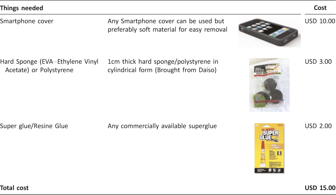
Table 1: Things needed to built a smartphone slit lamp camera adaptor

Portability

In comparison to the conventional slit lamp camera, the smartphone slit lamp camera is easily portable. It fits into a pocket and when it is needed, it can be easily mounted to an already existing slit lamp during routine examination.

Ease of use

Conventional slit lamp camera is usually placed in a special room and not easily accessible but with the smartphone slit lamp adaptor, it can easily be used in any slit lamp at anytime.

Patient will be explained of the anterior segment photography and consent will be taken.

Ease of Referrals

By using the smartphone slit lamp camera, pictures taken can be shared easily and securely to another colleague for further management or opinion. It can also be used by general practitioner who has a clinic equipped with a slit lamp.

How to DIY (do it yourself) a smartphone slit lamp camera

Determine the focal point of your smartphone by placing the camera aperture directly opposite the eye piece of the slit lamp making sure that placement is centred. The distance between your smartphone and the eye piece of slit lamp will be the focal length of your smartphone. This determines the thickness of the sponge you will be using for sponge Section B (Refer to Figure 1). The focal length for iPhone 4,4s,5, and 5c and 5s is 1.0cm and the focal length Samsung Galaxy Note I, II and III is 0.75cm. Figure 2 shows a completed DIY Smartphone Slit Lamp Adaptor.
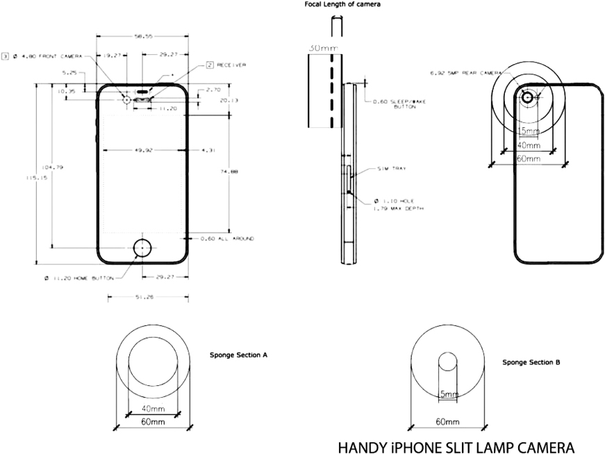
Figure 1: Blueprint of smartphone slitlamp camera
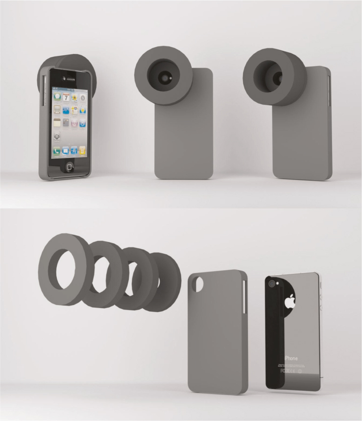
Figure 2: Smartphone Slit Lamp Camera

Prepare materials of sponges, super glue and surgical knife (Refer Figure 3, Picture 1). To make Section A sponge (Mounting of slit lamp), 1cm thick hard sponge is first measured by removing the eye piece from the slit lamp (Refer Figure 3, Picture No. 2). Use hard sponges or polystyrene that are 20mm wider than the eye piece so that the rim of the sponge is 10mm wide (Refer Figure 1, Sponge Section A). The author suggest to use Ethylene Vinyl Acetate (EVA) material as it is firm yet does not damage the slit lamp eye piece. Using the eye piece as a guide, a line is drawn using a pen for Section A – use the slit lamp eye piece (Refer Figure 3, Picture No. 2).
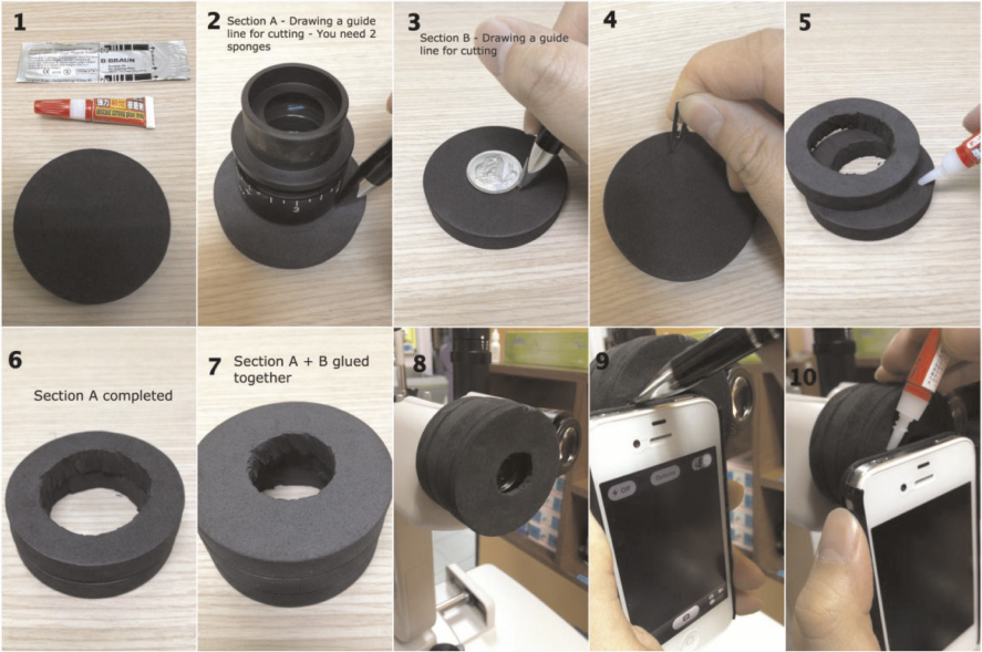
Figure 3: Steps of preparation of smartphone slitlamp adaptor

To make Section B sponge(the focal length of your smartphone), another 1cm thick hard sponge is used and measured with a small coin large enough for the smartphone camera hole (Figure 3, Picture No. 3). Caution needs to be taken during the measuring of the sponge making sure that the eye piece is in the centre.

All the sponges measured are then cut-to-fit the slit lamp eye piece and the viewing hole of smartphone camera (Refer Figure 3, Picture No. 4).

The sponges are then glued together using super glue using two to three pieces of sponge to form Section A and one piece of sponge to form Section B (Refer Figure 3, Picture No. 5–7). Attach the glued sponge to slit lamp after it is dry (Refer Figure 3, Picture No. 8).

The combined sponges are then glued to the smartphone cover by first marking the sponge (Refer Figure 3, Picture No.9). The smartphone is then inserted into the smartphone casing and then aligned with the slit lamp eye piece. Minimal amount of super glue is to be applied on the sponge to avoid spillage (Refer Figure 3, Picture No.10). Care is to be taken to ensure that the viewing hole is centred otherwise the end product will not be well aligned. All smartphones are generally suitable for mounting to the slit lamp (Refer Figure 4).
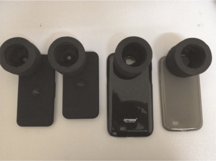
Figure 4: The finished products with different types of smartphones

The smartphone camera adaptor is then mounted to slit lamp and ready to be used. With the help of a VGA or HDMI cable of the smartphone (available commercially), we may also display the photo taken through a LCD projector, monitor or even an LED/LCD TV. Not only it can show live pictures but also the possibility of capturing videos and replay it and can be shown to the patient (Refer Figure 5).
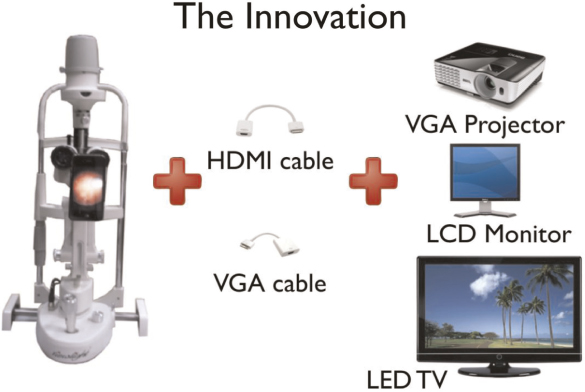
Figure 5: An innovative way to display anterior segment pictures through LCD projector, monitor or LED/LCD TV

How to use the smartphone slit lamp camera

After mounting the adaptor to the slit lamp camera, one may use the built-in camera app in your smartphone and just snap pictures or video as desired. The camera flash should be disabled.

Zooming in/out: It is recommended to use the slit lamp magnification for zooming in and out instead of the camera app as the quality of picture may reduce (Refer Figure 6).
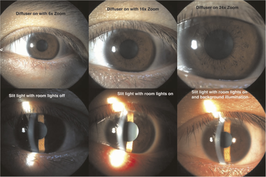
Figure 6: Different lighting and zooming of anterior segment pictures

Background lighting: It is recommended to switch on the room lights during picture taking. The quality of pictures improve with some additional background lighting (Refer Figure 6).

Diffuser: Some slit lamps comes with a diffuser which can be used to diffuse light if the ophthalmologist wishes to take pictures of the entire eye without slitting the light source. This gives a diffuse lighting to the eye. Refer to Figure 6 for detail.

AE/AF Lock: The picture quality may be increased by controlling lighting of the smartphone camera manually. AE (Auto Exposure) and AF (Auto Focus) lock enable the ophthalmologist to lock the exposure and focus to only on specific locations and lighting needs.

Quality of the pictures are comparable to the commercially available anterior segment camera (Refer Figure 7 & 8).
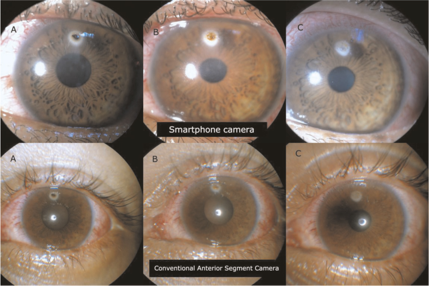
Figure 7: Sample pictures showing head to head comparison between Smartphone Camera (Top pictures) and conventional anterior segment camera (bottom) in a 20 year old patient with corneal foreign body. Picture A shows initial presentation. Picture B shows post-removal of corneal foreign body with retained rust ring. Picture C shows post removal of rust ring with corneal scar
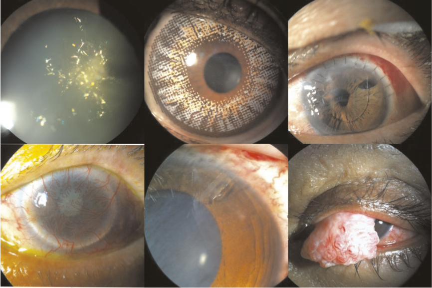
Figure 8: Samples of pitures taken with DIY – Smartphone Slit Lamp Camera

Conclusion

In the era today, most doctors are equipped with a smartphone which can help us not only in our daily lives but also in our work. Smartphone photography is something simple and yet very useful in the world of ophthalmology.

Though there might be concern regarding patient’s confidentiality when pictures taken are stored in the doctor’s personal smartphone, these problems may be solved by archiving the picture in a separate system like Picture Archiving and Communication System (PACS)7.

The use of smartphone photography in medical practice is not an uncommon practice. With this DIY guide, smartphone slit lamp anterior segment camera should no longer be seen as something unattainable to ophthalmologist. Instead it should be viewed as an indispensable accessory to slit lamp examination.

Acknowledgements

We greatly appreciate Dr Siti Zaleha Mohd Salleh, Dr Haireen Kamaruddin, Dr Azura Ramlee, Dr Nik Nazihah Binti Nik Azis, Mr Zawawi Zakaria, Ms Maziatul Nor Akmar for their contribution in the creation of iPhone slit lamp cameras.

References

1. Chhablani J, Kaja S, Shah VA. Smartphones in ophthalmology. Indian J Ophthalmol. Mar-Apr 2012;60(2):127–31.

2. Lord RK, Shah VA, San Filippo AN, Krishna R. Novel uses of smartphones in ophthalmology. Ophthalmology. Jun 2010;117(6):1274–1274 e1273.

3. Stanzel BV, Meyer CH. [Smartphones in ophthalmology: Relief or toys for physicians?]. Ophthalmologe. Jan 2012;109(1):8–20.

4. Tahiri Joutei Hassani R, El Sanharawi M, Dupont- Monod S, Baudouin C. [Smartphones in ophthalmology]. J Fr Ophtalmol. Jun 2013;36(6):499–525.

5. Bastawrous A, Cheeseman RC, Kumar A. iPhones for eye surgeons. Eye (Lond). Mar 2012;26(3):343–54.

6. Hester CC. Smart Phoneography – How to take slit lamp photographs with an iPhone. http://eyewiki.aao.org/Smart_Phoneography_-_How_to_take_slit_lamp_ photographs_with_an_iPhone. Accessed 21 September 2013.

7. Picture archiving and communication system (PACS). http://en.wikipedia.org/wiki/Picture_archiving_and_communication_system. Accessed 22 September 2013.

Read More

Posted on Feb 28, 2014 in Original Article | 9 comments

3D Printed Smartphone Indirect Lens Adapter for Rapid, High Quality Retinal Imaging

David Myung, MD, PhD1, Alexandre Jais, MS1, Lingmin He. MD, MS1, Mark S. Blumenkranz, MD1, Robert T. Chang, MD1

1Byers Eye Institute at Stanford, Stanford University School of Medicine, Stanford, California

Corresponding Author: viroptic@gmail.com

Journal MTM 3:1:9–15, 2014

doi:10.7309/jmtm.3.1.3


Obtaining a secure, photographic record of clinical findings during patient encounters can serve as a powerful adjunct to the otherwise text-heavy documentation that dominates modern electronic health record systems. This is particularly true in ophthalmology, which is one of several medical specialties that relies heavily on images for diagnosis and treatment. Conventionally, ophthalmic imaging has required expensive, tabletop units operated by a trained technician in an outpatient clinic setting. The ubiquity and evolution of smartphones into both high-resolution cameras and conduits for encrypted data transfer has the potential to change this; however, their use is inherently limited by the optics and lighting required to image the eye, and in particular, the retina. Here, we report the development of a lightweight, compact, user-friendly, 3D printed attachment enabling high quality fundus photos by coupling smartphones to indirect ophthalmoscopy condensing lenses. The attachment is designed to hold a specific lens at a prescribed but adjustable distance from the camera lens, can utilize either the phone’s native flash for lighting or another coaxial light source, and has the potential to be operated with one hand. Using both mechanical prototypes and subsequent 3D printed versions of the device, we were able to photodocument a variety of both normal and abnormal retinal findings.


Read More