Jan Bond Chan, (MBBS)1,2, Hao Chi Ho, (MBBS)1,2, Nor Fariza Ngah, (MBBS, MS Ophthal)1, Elias Hussein, (MBBS, MS Ophthal)1
1Department of Ophthalmology, Hospital Selayang, Malaysia; 2Department of Ophthalmology, Hospital Universiti Sains Malaysia
Corresponding Author: janbond@hotmail.com
Journal MTM 3:1:16–22, 2014
doi:10.7309/jmtm.3.1.4
Online Appendix
http://www.youtube.com/watch?v=jwq7nDwgxa0&feature=youtu.be
Video 1: Video of manufacturing and assembly of the device with example of usage.
Link to get raw material
http://www.daisojapan.com/p-20209-eva-cushion-d315-in-4-pcs-12pks.aspx
Introduction
In the Ophthalmology setting, taking pictures of the anterior segment is of crucial use for ophthalmologists in their daily practise. Ophthalmologists relies heavily of anterior segment camera for recording of progression of disease, documentation for case presentation and discussion, referrals to colleagues or subspecialty clinic and education for junior doctors and patients.
A conventional slit lamp camera is usually used for taking good quality pictures of the eye. The problem with slit lamp camera is that it is expensive and usually immobile.
The use of the smartphone in ophthalmology is more common than ever before1–5. Although most ophthalmology clinics are equipped with slit lamp but not all clinics are fortunate enough to be equipped with a high quality anterior segment slit lamp camera. Smartphones are usually incorporated with high resolution cameras which are commonly used by ophthalmologist capture pictures of the anterior segment of the eye 1–5. There are several commercially available smartphone adaptors to a slitlamp but they are expensive and is specific for certain slit lamp brands only6. We are writing to introduce an easy method to produce and use your own smartphone as a slitlamp anterior segment camera – Do it yourself (DIY).
Advantages of smartphone slit lamp camera
Cost saving and maintenance
Conventional slit lamp camera is expensive costing at about USD 15,000.00. The commercially available smartphone slit lamp camera adaptor (eg. EyePhotoDoc, Zarf iPhone Adaptor, Magnifi iPhone adaptor, Steady iPix Telescope Photo adaptor for iPhone, Keeler portable slit lamp, iExaminer, Tiger Lens and Skylight) ranges from USD 75.00 to USD 520.00. The estimated cost for a DIY smartphone slit-lamp adaptor is USD 15.00 (Refer Table 1) which reduces the cost significantly. A conventional slit lamp camera needs to be serviced and maintenance needs to be done which incurs additional cost to the ophthalmologist. By using the smartphone slit lamp camera, there is virtually zero maintenance.
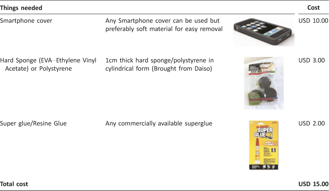
Table 1: Things needed to built a smartphone slit lamp camera adaptor
Portability
In comparison to the conventional slit lamp camera, the smartphone slit lamp camera is easily portable. It fits into a pocket and when it is needed, it can be easily mounted to an already existing slit lamp during routine examination.
Ease of use
Conventional slit lamp camera is usually placed in a special room and not easily accessible but with the smartphone slit lamp adaptor, it can easily be used in any slit lamp at anytime.
Patient will be explained of the anterior segment photography and consent will be taken.
Ease of Referrals
By using the smartphone slit lamp camera, pictures taken can be shared easily and securely to another colleague for further management or opinion. It can also be used by general practitioner who has a clinic equipped with a slit lamp.
How to DIY (do it yourself) a smartphone slit lamp camera
Determine the focal point of your smartphone by placing the camera aperture directly opposite the eye piece of the slit lamp making sure that placement is centred. The distance between your smartphone and the eye piece of slit lamp will be the focal length of your smartphone. This determines the thickness of the sponge you will be using for sponge Section B (Refer to Figure 1). The focal length for iPhone 4,4s,5, and 5c and 5s is 1.0cm and the focal length Samsung Galaxy Note I, II and III is 0.75cm. Figure 2 shows a completed DIY Smartphone Slit Lamp Adaptor.
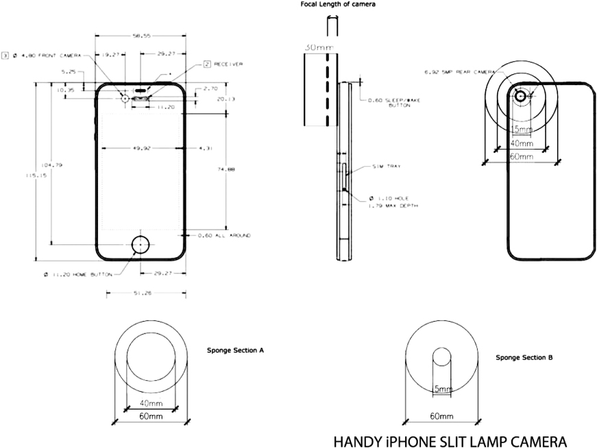
Figure 1: Blueprint of smartphone slitlamp camera
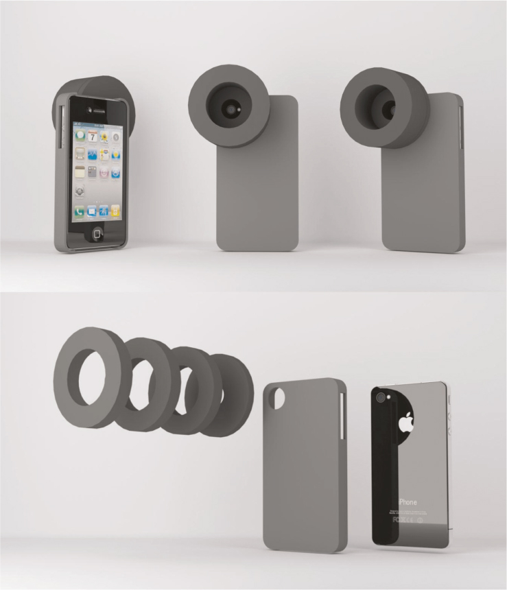
Figure 2: Smartphone Slit Lamp Camera
Prepare materials of sponges, super glue and surgical knife (Refer Figure 3, Picture 1). To make Section A sponge (Mounting of slit lamp), 1cm thick hard sponge is first measured by removing the eye piece from the slit lamp (Refer Figure 3, Picture No. 2). Use hard sponges or polystyrene that are 20mm wider than the eye piece so that the rim of the sponge is 10mm wide (Refer Figure 1, Sponge Section A). The author suggest to use Ethylene Vinyl Acetate (EVA) material as it is firm yet does not damage the slit lamp eye piece. Using the eye piece as a guide, a line is drawn using a pen for Section A – use the slit lamp eye piece (Refer Figure 3, Picture No. 2).
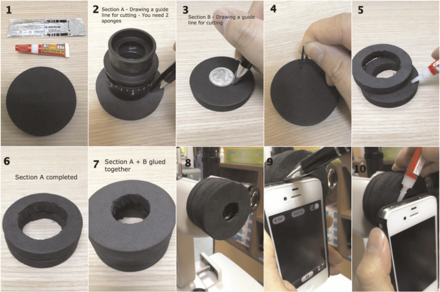
Figure 3: Steps of preparation of smartphone slitlamp adaptor
To make Section B sponge(the focal length of your smartphone), another 1cm thick hard sponge is used and measured with a small coin large enough for the smartphone camera hole (Figure 3, Picture No. 3). Caution needs to be taken during the measuring of the sponge making sure that the eye piece is in the centre.
All the sponges measured are then cut-to-fit the slit lamp eye piece and the viewing hole of smartphone camera (Refer Figure 3, Picture No. 4).
The sponges are then glued together using super glue using two to three pieces of sponge to form Section A and one piece of sponge to form Section B (Refer Figure 3, Picture No. 5–7). Attach the glued sponge to slit lamp after it is dry (Refer Figure 3, Picture No. 8).
The combined sponges are then glued to the smartphone cover by first marking the sponge (Refer Figure 3, Picture No.9). The smartphone is then inserted into the smartphone casing and then aligned with the slit lamp eye piece. Minimal amount of super glue is to be applied on the sponge to avoid spillage (Refer Figure 3, Picture No.10). Care is to be taken to ensure that the viewing hole is centred otherwise the end product will not be well aligned. All smartphones are generally suitable for mounting to the slit lamp (Refer Figure 4).
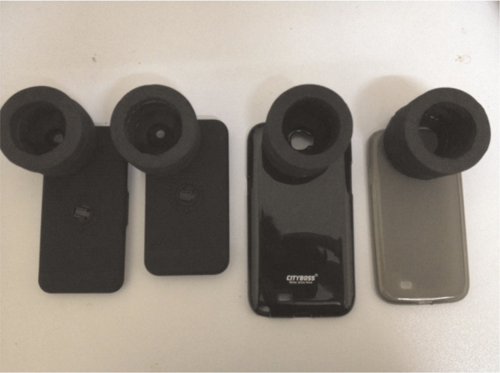
Figure 4: The finished products with different types of smartphones
The smartphone camera adaptor is then mounted to slit lamp and ready to be used. With the help of a VGA or HDMI cable of the smartphone (available commercially), we may also display the photo taken through a LCD projector, monitor or even an LED/LCD TV. Not only it can show live pictures but also the possibility of capturing videos and replay it and can be shown to the patient (Refer Figure 5).
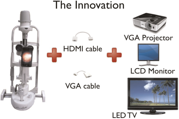
Figure 5: An innovative way to display anterior segment pictures through LCD projector, monitor or LED/LCD TV
How to use the smartphone slit lamp camera
After mounting the adaptor to the slit lamp camera, one may use the built-in camera app in your smartphone and just snap pictures or video as desired. The camera flash should be disabled.
Zooming in/out: It is recommended to use the slit lamp magnification for zooming in and out instead of the camera app as the quality of picture may reduce (Refer Figure 6).
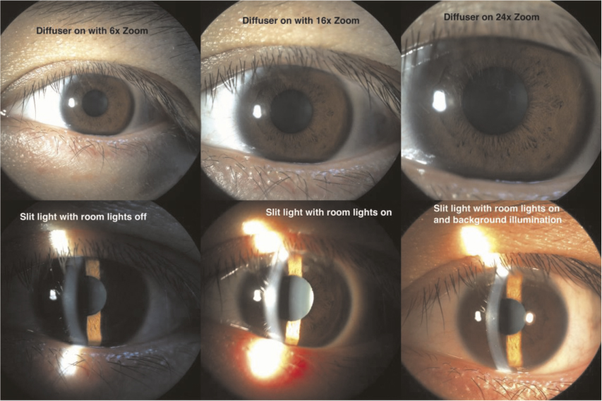
Figure 6: Different lighting and zooming of anterior segment pictures
Background lighting: It is recommended to switch on the room lights during picture taking. The quality of pictures improve with some additional background lighting (Refer Figure 6).
Diffuser: Some slit lamps comes with a diffuser which can be used to diffuse light if the ophthalmologist wishes to take pictures of the entire eye without slitting the light source. This gives a diffuse lighting to the eye. Refer to Figure 6 for detail.
AE/AF Lock: The picture quality may be increased by controlling lighting of the smartphone camera manually. AE (Auto Exposure) and AF (Auto Focus) lock enable the ophthalmologist to lock the exposure and focus to only on specific locations and lighting needs.
Quality of the pictures are comparable to the commercially available anterior segment camera (Refer Figure 7 & 8).
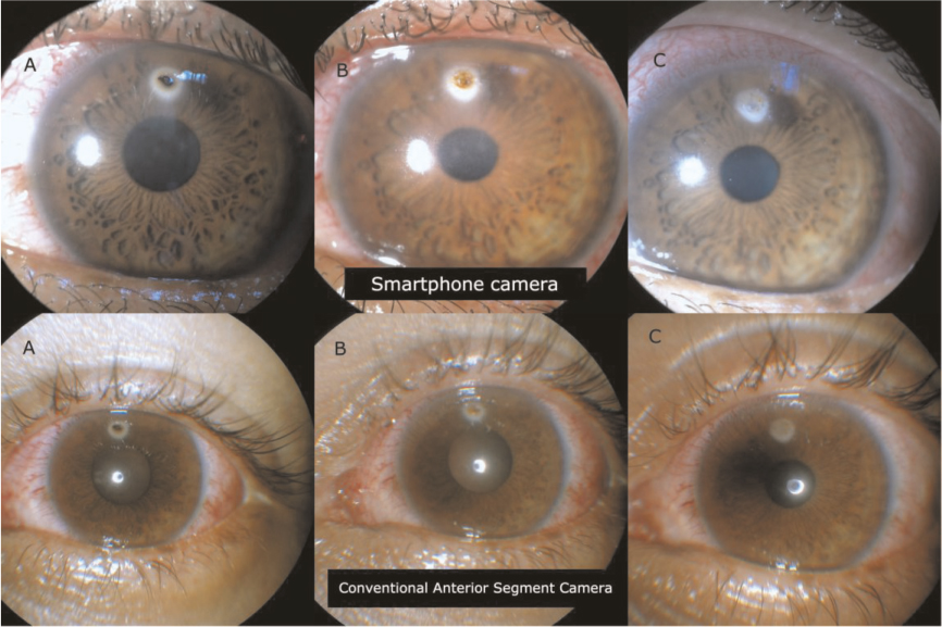
Figure 7: Sample pictures showing head to head comparison between Smartphone Camera (Top pictures) and conventional anterior segment camera (bottom) in a 20 year old patient with corneal foreign body. Picture A shows initial presentation. Picture B shows post-removal of corneal foreign body with retained rust ring. Picture C shows post removal of rust ring with corneal scar
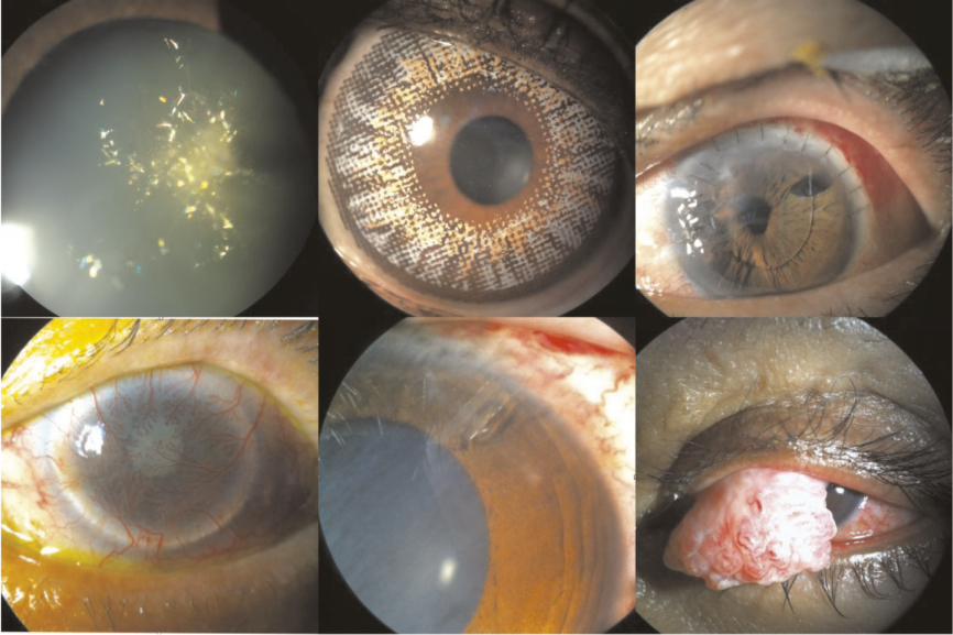
Figure 8: Samples of pitures taken with DIY – Smartphone Slit Lamp Camera
Conclusion
In the era today, most doctors are equipped with a smartphone which can help us not only in our daily lives but also in our work. Smartphone photography is something simple and yet very useful in the world of ophthalmology.
Though there might be concern regarding patient’s confidentiality when pictures taken are stored in the doctor’s personal smartphone, these problems may be solved by archiving the picture in a separate system like Picture Archiving and Communication System (PACS)7.
The use of smartphone photography in medical practice is not an uncommon practice. With this DIY guide, smartphone slit lamp anterior segment camera should no longer be seen as something unattainable to ophthalmologist. Instead it should be viewed as an indispensable accessory to slit lamp examination.
Acknowledgements
We greatly appreciate Dr Siti Zaleha Mohd Salleh, Dr Haireen Kamaruddin, Dr Azura Ramlee, Dr Nik Nazihah Binti Nik Azis, Mr Zawawi Zakaria, Ms Maziatul Nor Akmar for their contribution in the creation of iPhone slit lamp cameras.
References
1. Chhablani J, Kaja S, Shah VA. Smartphones in ophthalmology. Indian J Ophthalmol. Mar-Apr 2012;60(2):127–31. 
2. Lord RK, Shah VA, San Filippo AN, Krishna R. Novel uses of smartphones in ophthalmology. Ophthalmology. Jun 2010;117(6):1274–1274 e1273.
3. Stanzel BV, Meyer CH. [Smartphones in ophthalmology: Relief or toys for physicians?]. Ophthalmologe. Jan 2012;109(1):8–20.
4. Tahiri Joutei Hassani R, El Sanharawi M, Dupont- Monod S, Baudouin C. [Smartphones in ophthalmology]. J Fr Ophtalmol. Jun 2013;36(6):499–525. 
5. Bastawrous A, Cheeseman RC, Kumar A. iPhones for eye surgeons. Eye (Lond). Mar 2012;26(3):343–54.
6. Hester CC. Smart Phoneography – How to take slit lamp photographs with an iPhone. http://eyewiki.aao.org/Smart_Phoneography_-_How_to_take_slit_lamp_ photographs_with_an_iPhone. Accessed 21 September 2013.
7. Picture archiving and communication system (PACS). http://en.wikipedia.org/wiki/Picture_archiving_and_communication_system. Accessed 22 September 2013.
Read More
![]()
![]()


