Android Smartphone as an Alternative to Operating Microscope Camera for Recording High Definition Surgical Videos: Setup and Results.
Prof. Sanjiv Kumar Gupta1, Dr. Ajai Kumar2, Dr. Arun Sharma1, Dr. Siddharth Agrawal1, Dr. Vishal Katiyar1
1Department of Ophthalmology, King George’s Medical University, Lucknow, U.P., India; 2Jan Kalyan Eye Hospital, A-1040, Indira Nagar, Lucknow, U.P., India
Corresponding Author: sanjiv204@gmail.com
Journal MTM 4:3:39–42, 2015
Video recording and still photography is an essential component for documenting surgical and clinical details. Additionally videos have important role in skill transfer, demonstration of new procedures, and as material of clinical evidence. We here describe the use of an Android smartphone, (HTC Incredible S) for capturing High Definition (HD) video of ocular surgery through the assistant observer scope of an operating microscope.
We have described the arrangement used to mount the smartphone to microscope and discussed the advantages and limitations of this arrangement when compared to a conventional capturing and recording system used with the operating microscope.
Introduction
Video recording and still photography is an essential component of documenting surgical and clinical details. Videos have important role in skill transfer and training,1,2 demonstration of new procedures,3 and as material of clinical evidence. Conventionally the video and still imaging of surgical videos has been done using CCD (Charged Couple Device) camera attached to a beam splitter and C-mount. These cameras produce analog signal which is then transferred to a recording device (VCR, DVD recorder, Hard Disk Recorder, Commuter video Capture Card etc.). Later on this is edited using computer to produce a video of desired quality, with narration and captions.
Recently the availability of smartphones with processing capabilities comparable to computers and plethora of sensors has made their application in medical science increasingly popular.4 The camera interface of these smartphones has rich features and has numerous advantages over the conventional imaging system used in ophthalmology, both for stills and videos. Further we will be discussing only the smartphones with Android operating system and camera supporting at least 8 megapixel camera and full HD (1280*720 pixels) video recording at 30 frames per second.
We here describe the use of one such smartphone, (HTC Incredible S) for acquiring High Definition video of ocular surgery through standard operating microscope with assistant observer scope.
This kind of arrangement can be cost effective and capture high quality images and videos with added advantages of easy sharing and transfer to other devices.

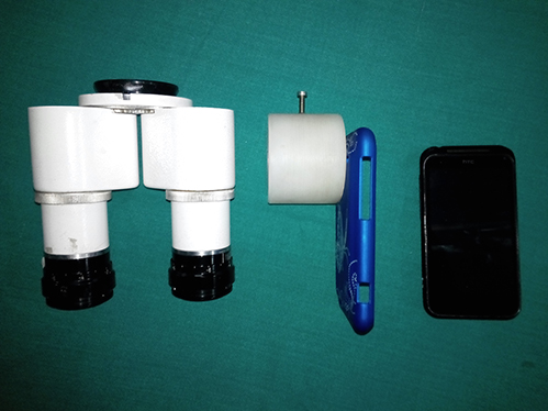
Figure 1: The components for attaching the smartphone to the assistant viewing scope from right to left, The assistant binocular scope detached from the operating microscope, Smart phone case with attached tube for connecting it to the assistant scope, The smart phone.
Subjects and Methods
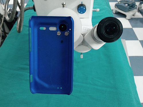
An operating microscope (Model YZ20T, from 66 Vision-Tech Co., Ltd, China), with binocular assistant observation tube was used in conjunction with an android Smartphone (HTC Incredible S). The phone has Android operating system 4.1 version with 8 megapixel camera with autofocus and full HD (High Definition 1280*720) video capturing capability. An adaptor was developed to hold the phone securely in place aligned to the optics of the assistant viewing scope with help of supporting engineering staff (L & M Automatics, H-3 Panki Industrial area, Site-1, Kanpur, U.P., India. landmautomatics@gmail.com). The phone was cradled in a hard phone case compatible for the phone model. In turn this phone case was mounted to an adaptor which was secured and optically aligned to one of the oculus of the assistant observation scope. The components and the assembled arrangement are shown in the photographs.

Figure 2: Smart phone case mounted on the assistant viewing scope
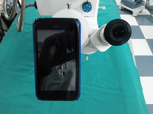
Once assembled, the camera software on the phone was turned on and the microscope was focussed and centred on the operating field. The phone screen showed the view congruent to the surgeon’s view and the independent focus and magnification control of the observer tube was used to obtain focus and desired level of field/zoom in the phone screen. Fine focus was done using touch to focus feature of the camera software of the phone. This also helped in the attaining proper white balance and exposure control of the area of interest in the phone’s camera viewfinder.

Figure 3: The complete assembly of smart phone mounted on the assistant viewing scope, ready to capture images and videos.

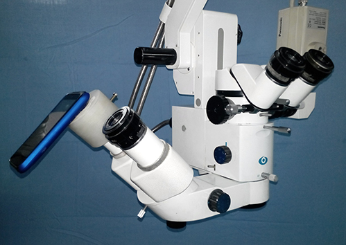
Figure 4: The complete setup showing the microscope optical head with the attachment to record images and videos using smart phone.
This arrangement allowed full HD video capturing and 8 Megapixel (3264*2448 pixels) still photos. The arrangement allows simple and intuitive touch based control of still image capturing, video recording start and stop process, and touch to focus the area of interest on the viewfinder screen. The video and stills are saved on the device removable 16 Gigabyte memory card which can be easily accessed using USB (Universal serial Bus) cable attached to computer.
Results
The captured videos had to be rotated 90 degree to obtain surgeon’s perspective view which is used as conventional viewing angle for surgical videos.
The captured videos and still show excellent clarity and being full HD can be viewed in original resolution without any pixilation when viewed on computer, TV or projected through a projected. These videos were better than the standard definition videos captured using the pre-existing CCD camera with DVD recording system.
The still images captured also needed to be rotated but apart from that they were excellent in sharpness, resolution and colour saturation.
Discussion
In the present era of evidence based medicine videos and photographs make an essential component of documenting the surgical events and cases. The video presentations also aid in skill transfer which otherwise is not possible using still photographs and text. Importance of a good video in scientific presentations to draw the attention of the audience and make them understand the steps of the procedure cannot be overemphasised.
Present era arrangement of video capturing and recording consists of beam splitter, C mount, a CCD camera (Standard definition or High Definition), a display system (TV or computer screen), a recording system (DVD recorder, Hard disk based recorder or computer attached video capture cards) and significant lengths of electric and video cables. This arrangement cause’s loss of quality of video signal and due to its nature is prone to frequent breakdown.
The smart phone based solution is single component system incorporating capturing, recording and viewing system attached to the microscope. The video quality can be easily controlled using the phone software which includes the focus, exposure control, colour hue, coloursaturation and the resolution of the video.
The arrangement is especially useful for capturing posterior segment video and images which suffer from overexposure due to very bright operating field surrounded by the unlit dark area. This arrangement with a touch screen allows touch to focus/exposure setting to easily focus and adjust the exposure according to the illumination of the area of interest.
The additional software on the smartphone allows for immediate review of the recorded video on the device screen. Limited editing of the still images and video is also possible on the device itself.
The recorded video does not capture any other detail apart from the view obtained through the operating microscope view hence unless the patient’s identity is manually added to the file details the patients’ privacy is not compromised.
Additionally sharing the video online is possible by attaching a video out cable from the phone to any television with HDMI input. Offlinesharing is possible through Wi-Fi, Bluetooth or USB connectivity, out of which USB may be the preferable as the video files are large in size and thus its high transfer speed will be faster. There is no need of burning DVDs or CDs as may be the case with Computer or DVD recorder based recording.
The arrangement of using smartphone camera for capturing, recording video and still images is a single device solution which is inherently simple cost effective, easily available, robust system which is less likely to fail. The videos and images are in full high definition resolution which can show the minute details of surgical procedures. Additionally image quality adjustments and easy transfer of the video are features which are not possible with conventional recording system.
This arrangement occupies one of the oculus of the assistant scope and hence the assistant cannot get the same conventional stereoscopic view needed for ideal assistance. Though this limitation can be overcome by mounting the phone through an appropriate beam splitter.
The recording time will be limited by the memory space of the micro SD card used. Presently these memory cards are available in sizes from 8 Gigabyte to 128 Gigabyte allowing recording duration of 1 hour to 16 hours of HD video. This limits the amount of video which can be stored in the device and will need frequent transfer to a device with higher storage capacity. This issue can be solved with the new generation of phones having capability to support external storage device using OTG (On The Go) cable attached to the micro USB port of the device. Thus not only this will increase the storage space available for recording but also make the transfer of the recorded video fast and easy. Further this arrangement presently does not allow capture of 3D images or videos.
In conclusion this arrangement of using Android smartphone to capture images and high definition video through the assistant scope of the operating microscope is compact, robust with easy storage, preview, sharing arrangements. The ease with which the exposure and focus control can be adjusted for optimum image and video is additional feature not possible with conventional CCD camera arrangement presently used.
Acknowledgements
None
References
1. Quellec G, Lamard M, Cochener B, Cazuguel G. Real-time segmentation and recognition of surgical tasks in cataract surgery videos. IEEE Trans Med Imaging. 2014;33:2352–60. ![]()
2. Gooi P1, Ahmed Y1, Ahmed II2. Use of a microscope-mounted wide-angle point of view camera to record optimal hand position in ocular surgery. J Cataract Refract Surg. 2014;40:1071–4. ![]()
3. Quellec G1, Charrière K2, Lamard M3, Droueche Z2, Roux C2, Cochener B4, Cazuguel G2. Real-time recognition of surgical tasks in eye surgery videos. Med Image Anal 2014;18:579–90. ![]()
4. Bastawrous A, Cheeseman RC, Kumar. A iPhones for eye surgeons. Eye 2012;26:343–54. ![]()

